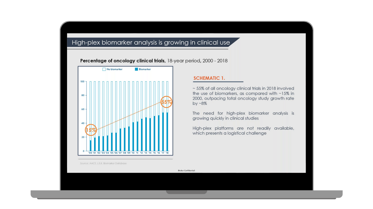
Pre- and post-treatment immunological profiling of both peripheral blood and the tumor microenvironment is a useful strategy to discover processes and biomarkers which might aid in developing and commercializing novel precision immunotherapies. ChipCytometry™ allows for the quantitative analysis of virtually unlimited protein biomarkers in a single sample. The technology combines high-resolution HDR imaging with AI-powered cell-segmentation software to deliver quantitative biomarker analysis with true single-cell resolution, enabling deep phenotyping of every individual cell in each sample.
With over 40 validated markers for tissue samples and more than 110 validated markers for PBMCs, this technology has broad applications to clinical and pre-clinical studies in oncology, immunology and drug development. ChipCytometry ™ clinical studies now offer CytoChex® collection tubes which minimize adverse effects of time, storage, processing, and shipping on sample integrity.
Identifying rare cell populations to better understand how they influence human health and development requires high-plex technologies, but unlike flow cytometry and RNA sequencing, ChipCytometry™ offers the spatial information necessary to interpret how rare cells are interacting with other cells in the microenvironment.
Hosted by The Scientist, this webinar features two researchers who explore the latest biological insights learned from studying rare cell populations in the nervous system.

Researchers are working to understand the impact of COVID-19 at the molecular level and how various biological processes are affected, leading to more-severe illness. In this 15-minute webinar, we will discuss how the nCounter® platform can be used in the development of tools and therapeutics against COVID-19. We will review some case studies utilizing the Host Response Panel to understand the role that autoantibodies against Type I IFN play in life-threatening COVID-19 illness.
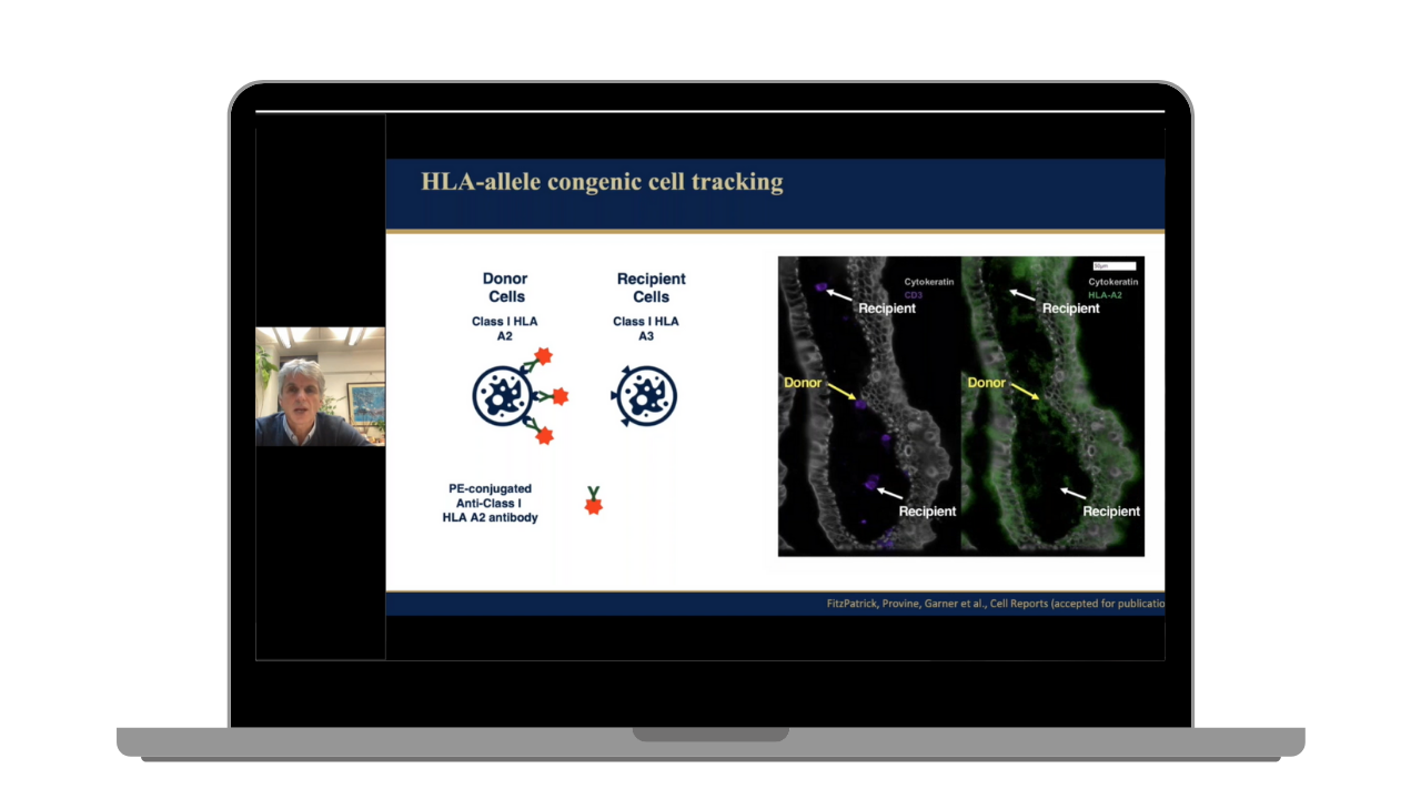
Paul Klenerman's research group has been exploring the role of human tissue resident immune cells in host defense, focusing on innate-like T cells such as MAIT cells. They have used ChipCytometry™ to explore the distribution of these cells in tissues such as the GI tract and also human lymphoid tissue. This webinar will present some data on recent findings and explore the biology as well as the technical aspects of studies of these tissue-associated populations in humans.
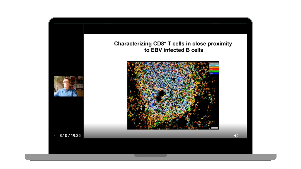
Christian Münz's research group has a long-standing interest in studying human lymphotropic viral infections and their immune control, including Epstein Barr virus (EBV). For this purpose, they study mice with reconstituted human immune system components (humanized mice) that can be infected, develop pathologies and mount cell mediated immune responses to these viruses. They have employed ChipCytometry™ to localize EBV infection in the spleen with and without compromising its immune control by blocking an important co-stimulatory molecule, namely CD27, that certain CD8+ T cells need to kill EBV infected B cells.
In this webinar, Christian will discuss localization of infected B cells and activated T cells in the white pulp of infected spleens, their co-stimulatory and co-inhibitory receptors, as well as markers of their differentiation and homing. They are aiming to employ ChipCytometry™ to gain spatio-temporal insights into the immune control of the most ubiquitous and most oncogenic human viral infection.
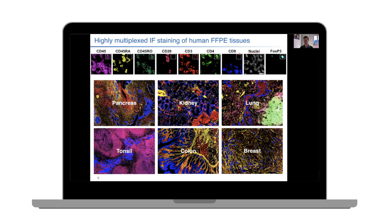
Simultaneous in-situ analysis of a multitude of cells and markers is important to our understanding of tissue biology in health and disease. ChipCytometry™ is a powerful tool for such analysis and can be used on both FF and FFPE tissue samples. In this webinar, we will describe the systematic optimization of high-plex staining and analysis of FFPE samples using ChipCytometry.
This webinar is part of our ChipCytometry™ User Webinar Series.
The recent shift from bulk data to individual cellular profiles unveiled how diverse cellular populations can be. The next challenge is to understand how these different individual cells interact with each other to create systems. In this webinar, researchers Timothy O’Sullivan and Jacob Blum will examine how they apply data from numerous individual cells within physiological and pathological contexts to better understand mechanisms of health and disease.

Multi-omics technologies are increasingly integral in neurobiology research for their ability to shed light on diverse cell types, cell states, cellular interactions, as well as their roles in CNS disorders.
Using multi-omics technologies can often be complex and intimidating so we have brought together a group of early-career scientists who are actively involved in using these diverse techniques to address neurobiology questions. These hands-on scientists are keen to share their knowledge and expertise in the preparation of cells and reagents, setting up assays and instruments, sorting and analyzing neural cells from various sample types, and much more.

Spatial biology helps researchers understand how location and proximity influence and modulate function at large within the context of physiological and pathological effects. In this webinar from The Scientist, Elana Fertig and Sanjay Srivatsan will discuss the importance of spatial data and how to extract insights from high-throughput datasets. They will cover machine learning for spatial multi-omics in cancer biology and embryo-scale spatial transcriptomics with single-cell resolution.
Canopy Biosciences' ChipCytometry™ platform is capable of extracting precise, spatially resolved, quantitative immunoprofiling data from clinical tissue samples. Clinical tissue specimens are largely prepared as formalin fixed, paraffin embedded (FFPE) blocks. FFPE presents unique challenges for those developing robust multiplexing assays. In this webinar, we discuss our strategies for dealing with three of these challenges: FFPE archived sample integrity, antigen retrieval or unmasking, and tissue autofluorescence.

In this webinar, learn about the value and utilization of NanoString® nCounter® panels and the GeoMx® Digital Spatial Profiler (DSP) for preclinical studies that integrate bulk gene expression and spatial biology to maximize insight from precious samples.

In this webinar, you'll hear from both Canopy and NanoString experts to learn more about NanoString’s GeoMx Digital Spatial Profiler (DSP).
GeoMx DSP combines the best of spatial and molecular profiling technologies by generating a whole tissue image at single cell resolution and digital profiling data for 10’s-1,000’s of RNA or Protein analytes from up to 12 tissue slides per day.
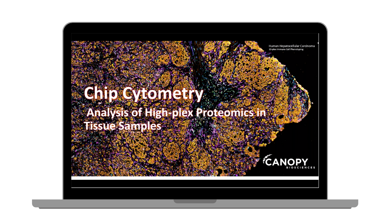
ChipCytometry™ is an image-based proteomics platform that allows for quantitative analysis of protein biomarkers in tissues with true single-cell resolution and spatial context. In this webinar, we will discuss the basics of ChipCytometry and demonstrate the powerful image processing capabilities of high-plex data sets in tumor samples.

Standard NGS methodology has an error rate of 1-3%, making the detection of low-frequency variants difficult to decipher from the sequencing noise. For researchers studying clonal hematopoiesis it is critical to have an increased limit of detection to see mutated clonal populations. Recent studies have indicated these variants are present below the threshold of traditional sequencing strategies. RareSeq from Canopy Biosciences has been shown to be successful in identifying these mutated clonal populations sooner, potentially leading to better patient outcomes.

Most biomarker analysis works in Phase I and II clinical trials are exploratory clinical research studies. The data will then guide future clinical development or plan subsequent studies. With the clinical impact of this work, it's hard to know what level of lab certification you need— CLIA? GLP? GCP? CAP?
In this 15-minute webinar, we will discuss the lab certifications out there and help navigate who needs what for their level of lab research.
The ChipCytometry™ platform incorporates a revolutionary sample receptacle device called a ZellSafe Chip. This unique technology enables simple and reliable sample preservation that eliminates cryo-bias and allows for re-interrogation of precious clinical samples. In this webinar, we will discuss the advantages of employing ChipCytometry in clinical trials.

With NanoString's digital counting technology, even highly degraded RNA yields high quality and highly reproducible results. FFPE is an extremely common sample—from research labs to clinical trials, there are FFPE samples everywhere. The challenge is how to analyze them. High variation, low yield, & high degradation makes them notoriously difficult. In this 15-minute webinar, learn how the robust nCounter gene expression assays have solved this issue allowing you to analyze up to 800 genes in a single sample.

In this 15-minute webinar, we will review the different types of ELISAs used for HCP detection in biologics. We will also give an overview on how to choose the best approach to find the right assay for your process.

In this 15-minute webinar, we will demonstrate the updated features of the ZellExplorer App, including new multi-gate display and hybrid scaling. Spencer Schwarz, Field Application Scientist, will give a live demo and answer any questions you might have about the new software features.

This 15-minute webinar discusses ChipCytometry™–a powerful image-based multiplex cytometry technology. Through the immobilization of cells or tissue samples on fluidic chips, the technique can be used to analyze tissue samples or phenotype individual cells. We explore the importance of resolution, dynamic range, and robust data analysis to generate actionable data from ChipCytometry platforms.

This 15-minute webinar explores Canopy Biosciences’ new interactive data analysis platform Rosalind. We provide an overview of the package we offer for data analysis of our NanoString + RNA-Seq services. Then we dive into a live demonstration of the interactive software so you can see its features highlighted.
This 11-minute webinar discusses our NanoString high throughput gene expression analysis service. The system is optimal for Oncology, Neuropathology, Infectious disease, and Immunological Research. NanoString works great with both fresh and FFPE tissue, blood, CSF, and isolated RNA. Receive a full data analysis report in a 2 week turn around time.
Canopy Biosciences is a world leader in multi-omics immune profiling technologies. With a global footprint, Canopy Biosciences offers a range of products and services that have helped many of the top companies and academic institutions further their clinical and translational research programs.